
After accounting for planned breach, the effective breach rate was 3.8% resulting in 96.2% accuracy for pedicle screw placement. Average screw insertion time was 1.76 ± 0.89 min. Among lateral breach (n = 16), ten screws were planned for in–out–in pedicle screw insertion. We noted 27 pedicle screw breach (medial: 10 lateral: 16 anterior: 1). We had 242 grade 2 pedicles, 133 grade 3, and 77 grade 4, and 44 pedicles were unfit for pedicle screw insertion. The average Cobb angle was 68.3° (range 60°–104°). Breach greater than 2 mm was considered for analysis. Analysis was performed to estimate the accuracy of screw placement and time for screw insertion.

Pedicles were classified according to complexity of insertion into five types. Materials and methodsģ1 complex spinal deformity correction surgeries were prospectively analyzed for performance of AIRO® mobile CT-based navigation system. To develop a classification based on the technical complexity encountered during pedicle screw insertion and to evaluate the performance of AIRO® CT navigation system based on this classification, in the clinical scenario of complex spinal deformity. Rajasekaran, Manindra Bhushan, Siddharth Aiyer, Rishi Kanna, Ajoy Prasad ShettyĪugust 2018, Volume 27, Issue 9, pp 2339 - 2347

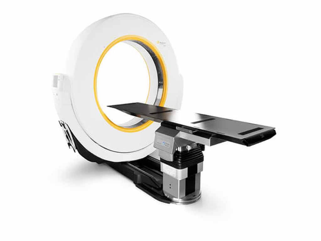
Weaver nor any member of his immediate family has financial interest in Mobius Imaging or Brainlab.S. "This tool is just another example of the leading edge surgical capabilities available to patients at Temple University Hospital."Įditor's Note: Neither Dr. "We are able to easily move this imaging tool, which allows us to give more patients access to this technology," added Dr. The device also offers added flexibility because it is small enough to fit through standard doors and moves easily around the hospital. This allows us to make rapid decisions and to verify optimal surgical results as the final step before closing up the patient."Īiro, developed by Mobius Imaging and distributed by Brainlab, connects to the Curve™ Image Guided Surgery System, a GPS-like navigation system for the human body, to further refine spatial positioning of the physician's surgical instruments.Īiro is designed to offer the surgical team unparalleled flexibility in positioning the patient and helps expand the use of intraoperative imaging for several surgical specialties, including neurosurgery and orthopaedic and trauma surgery. "Airo gives us access to intraoperative CT images of the patient's anatomy while they are still in the operating room. "Temple University Hospital is home to a world-class surgical team, and this new tool enhances our ability to provide our patients with top quality surgical care," said Michael Weaver, MD, Chair of the Department of Neurosurgery at Temple University School of Medicine.

Airo gives surgeons the ability to see real time, intraoperative (during surgery) anatomical detail, allowing for timely and informed decision-making. Temple University Hospital is the first hospital in the Philadelphia region to offer the Airo® Intraoperative CT (computerized tomography), an innovative imaging tool that provides diagnostic quality images and is designed for use in the operating room.


 0 kommentar(er)
